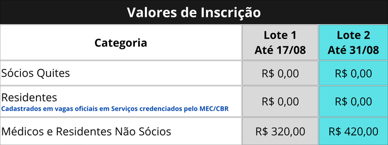
R$320,00
Format: Online – Live
The registration fee will be calculated by category and period (Batch).
Target Audience (Prerequisite):
Doctors and Residents in Radiology and Diagnostic Imaging
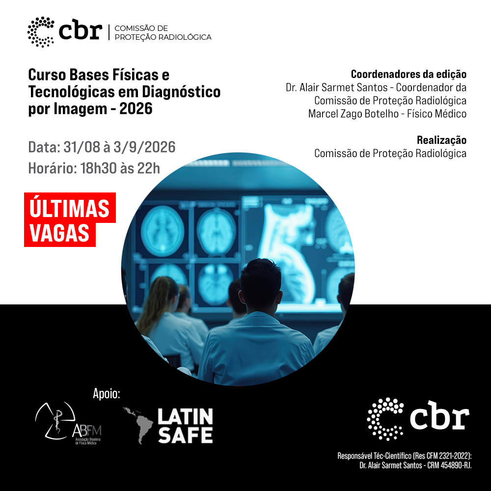
Coordination:
Dr. Alair Sarmet Santos
Physician Marcel Zago Botelho
Technical-Scientific Manager (CFM Resolution 2321-2022):
Dr. Alair Sarmet Santos – CRM 454890-RJ.
78 Vagas disponíveis
Description
“"Medical Physics for Physicians in Radiology and Diagnostic Imaging"”
BASIC BIBLIOGRAPHY
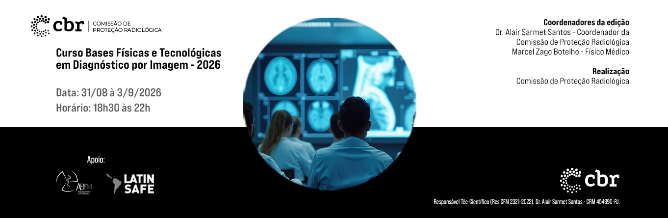
Physical and Technological Bases in Diagnostic Imaging
PRESENTATION
The Radiation Protection Committee of the Brazilian College of Radiology and Diagnostic Imaging (CPR/CBR) works to strengthen the culture of safety, technical qualification, and the responsible use of imaging technologies. Its educational agenda bridges the gap between the fundamentals of medical physics and clinical decisions that determine diagnostic quality, patient exposure, and the safety of healthcare teams.
The 2026 edition updates the course to a competency-based approach. Instead of presenting physics as isolated content, the program covers the entire examination chain—indication, acquisition, reconstruction, visualization, interpretation, and monitoring—demonstrating how technical choices modify contrast, resolution, noise, artifacts, and dose.
OBJECTIVE
Review and apply the physical, technological, and radiological protection fundamentals relevant to medical practice, using and disseminating the CBR/ABFM's "Physical and Technological Bases in Diagnostic Imaging" manual as the main reference for professional training and updating. The aim is to enhance the participant's ability to recognize limitations, optimize protocols, evaluate image quality, and participate in safety decisions in the main diagnostic imaging modalities.
METHODOLOGY
The course will be theoretical and intensive, with a scientific program focused on the practical application of radiodiagnostic physics and radiation protection concepts in the clinical context of different imaging diagnostic modalities. Classes will be expository and interactive, taught by a multidisciplinary team composed of medical physics professionals, radiodiagnostic physics specialists, and radiologists working in various areas of imaging diagnostics. The training will be conducted online via the Zoom Meeting platform, which offers interactive resources designed to encourage active student participation during the sessions. Mechanisms such as live chat, real-time feedback, a question-and-answer system with statistical visualization, and multiple-choice polls configured as questionnaires with the possibility of anonymous responses will be used. The results of these polls and questionnaires will be viewed in real time during the meeting and also exported as a report at the end of each session. This approach aims to stimulate participant engagement and encourage content retention, contributing to a more active and effective learning process.
PROGRAM CONTENT
THEMATIC AREA 1 | Physical and Technological Bases of X-rays
– Nature of radiation;
– Production and emission of X-rays;
– Spectrum;
Photoelectric and Compton interactions;
– Pipe components;
– Cathode, anode, and focal point;
Anodic effect;
– Generating systems;
– Filtration;
Collimation;
– Anti-scatter grids;
– Geometric factors and technical parameters related to image formation and quality.
THEMATIC AREA 2 | Fundamentals of Digital Radiology
– Digital image formation;
Matrix, pixel, bit, and grayscale;
– Response function and dynamic range;
– Computed radiography (CR) and digital radiography (DR);
– Direct and indirect conversion systems;
Acquisition, processing, visualization, and storage;
Spatial and contrast resolution;
Noise and signal-to-noise ratio;
– Exposure indicators;
Monitors and devices.
THEMATIC AREA 3 | Physical and Technological Bases Applied to Mammography
– Characteristics of breast tissue;
– Principles of mammographic image formation;
– X-ray equipment and tube;
– Emission spectrum and target/filter combinations;
– Automatic exposure control;
Breast compression;
– Anti-scatter grids and amplification techniques;
– Digital acquisition systems and detectors;
– Factors related to image quality and mean glandular dose;
– Artifacts;
– Concepts of tomosynthesis and fundamentals of quality control.
THEMATIC AREA 4 | Technological Bases of Fluoroscopy and Interventional Procedures
– Principles of fluoroscopic image formation;
– Image intensifiers and flat detectors;
– Automatic intensity control;
Collimation, filtration, field size, and magnification;
Continuous and pulsed fluoroscopy;
– Equipment settings;
Factors that affect image quality and exposure;
– Fluoroscopy time, reference air kerma, and kerma-area product;
– Basic concepts of patient and worker protection.
THEMATIC AREA 5 | Physical and Technological Bases of Computed Tomography
– Principles of tomographic image formation;
– Axial and helical acquisition;
– Attenuation coefficient and Hounsfield units;
Matrix, pixel, voxel, window, and level;
– Components of the CT scanner;
– Tubes, filters, collimators and detectors;
– Basic acquisition parameters, including kV, mAs, collimation, and pitch;
– Pre-processing and processing;
Filtered backprojection and notions of iterative reconstruction;
Factors related to noise, artifacts, image quality, and dose;
– Concepts of CTDIvol and DLP.
THEMATIC AREA 6 | Physical Principles of Magnetic Resonance Imaging and Ultrasound
In magnetic resonance imaging:
– Spin and magnetization;
– Precession and resonance;
Radiofrequency; T1 and T2 relaxations;
– Weighting and pulse sequences;
– Gradients, spatial encoding, and k-space;
– System components;
– Safety artifacts and fundamentals.
In ultrasound:
– Nature and propagation of sound;
Frequency and wavelength;
– Acoustic impedance;
Reflection, refraction, scattering, and attenuation;
– Transducers;
– Beam properties;
Spatial resolution;
– Profit and TGC;
– A, B, M and Doppler modes.
THEMATIC AREA 7 | Concepts of Radiological Protection, Standards and Quality Control
– Quantities and units in radiation protection;
– Biological effects;
– Principles of justification and optimization;
– Dose limitation for workers and the public;
– Diagnostic reference levels;
– Protection of patients, workers and the public;
– Radiation Protection Program;
Occupational monitoring and protective devices;
– Fundamentals of quality assurance and control;
– Image characteristics;
Acceptance and consistency tests;
Responsibilities;
– ANVISA Resolution No. 611/2022 and corresponding Normative Instructions.
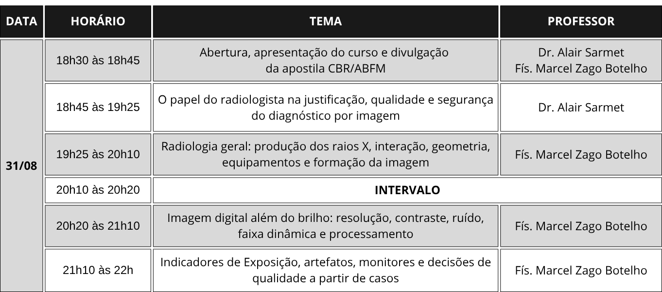
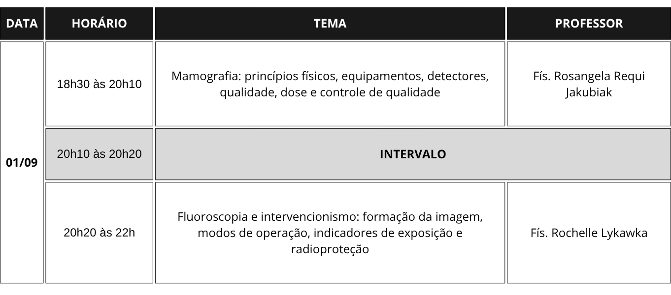
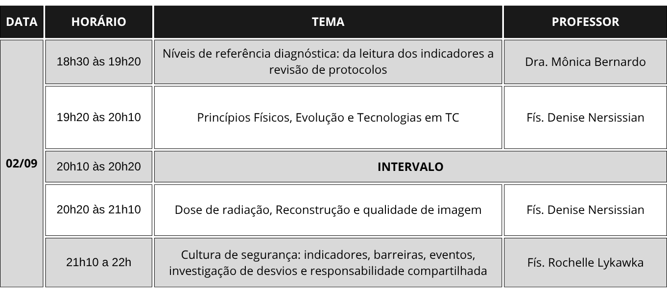
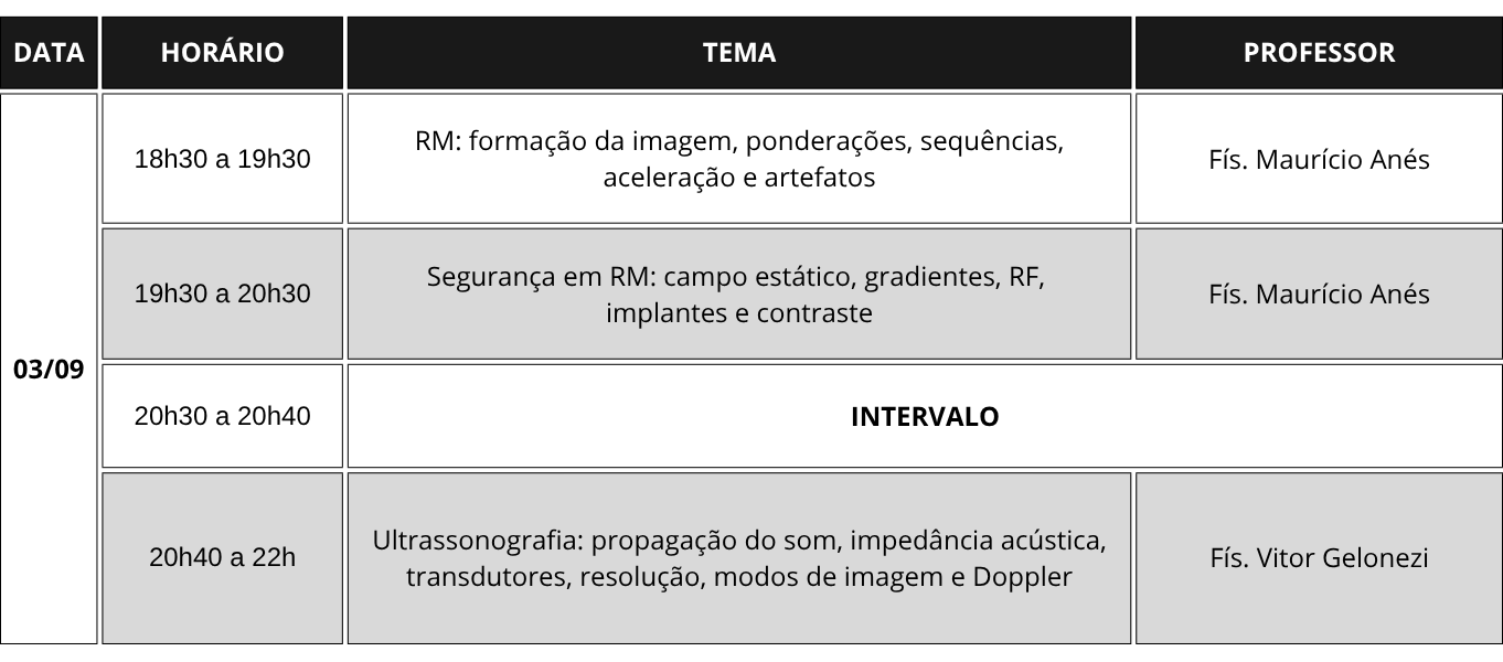
SCHEDULE
TEACHERS
Dr. Alair Sarmet
Coordinator of the CBR Radiological Protection Commission, Graduated in Medicine from the Fluminense Federal University-UFF, Master's Degree in Medicine (Radiology) from UFRJ (1992), PhD in Medicine (Radiology) from UFRJ (1996), Associate Professor and Head of the Rad. Dept. of the Faculty of Medicine, UFF, Full Member of ACAMERJ (Academy of Medicine of the State of RJ), Former President of CBR (2019-2020) and Member of the Advisory Board.
Dr. Monica Bernardo
Vice Coordinator of the CBR Radiological Protection Commission, Member of the CBR Criteria Adequacy Committee, President of Latin Safe, PhD in Sciences from the Faculty of Medical and Health Sciences at UNICAMP, Post-doctorate from the Institute of Physics of the University of São Paulo, Professor of the Radiology Knowledge Area at FCMS PUC SP/Sorocaba and Fellow University of Minnesota-USA.
Denise Nersissian
PhD in Nuclear Technology – Applications – IPEN/CNEN; Specialist in Radiodiagnosis from the Brazilian Association of Medical Physics (ABFM); Quality Control in Diagnostics by the Dosimetry Group Department of Radiation and Medical Physics at IF/USP; Preceptor of the Residency Program in Professional Health Area: Medical Physics – Radiodiagnosis area of the Faculty of Medicine at USP (University of São Paulo).
Rochelle Lykawaka
Medical Physics Specialist in Radiodiagnostic Physics ABFM, Specialist in Risk Management and Hospital Safety, Master in Medical Sciences UFRGS, Coordinator of the Radiological Protection Commission of HCPA/UFRGS, Director of the Radiodiagnostic Area ABFM and Member of the CBR Radiological Protection Commission.
Marcel Zago
Bachelor of Physics with a Specialization in Medical Physics – UFN/RS, Master's Degree in Nanosciences – UFN/RS, Specialist in Radiodiagnostic Physics by – ABFM-RX-288/1107, Technical Director of STAFF – Solutions in Medical Physics and Radiation Protection, Member of the CBR Radiological Protection Commission, Member of the Radiological Protection Commission of the SBP and Former President of the Brazilian Association of Medical Physics – ABFM.
Mauricio Anés
Graduated in Physics from the Pontifical Catholic University of Rio Grande do Sul (2000), he holds a degree in Radiodiagnostic Physics and Nuclear Medicine from the Brazilian Association of Medical Physics (ABFM). He is a full member of the ABFM and ISMRM (International Society of Magnetic Resonance in Medicine). He is currently a physicist at the Hospital de Clínicas de Porto Alegre, providing advisory and consulting services in safety and quality in diagnostic and interventional radiology. He has experience in Medical Physics, with an emphasis on Medical Radiology, focusing primarily on the following topics: MRI and neuroimaging.
Vitor Gelonezi
Medical Physicist specializing in Radiodiagnosis and Nuclear Medicine; Radiation Protection Supervisor in Nuclear Medicine certified by CNEN; Technical Consultant at Metrobras – Radiation Metrology and Personal Dosimetry; Managing partner of KEV – Diagnostic Imaging Consulting; ISO 17025 Auditor; Internal Auditor PADI-CBR.
Rosangela Jakubiak
Graduated in Physics from the Federal University of Paraná, Master's and PhD in Biomedical Engineering and Industrial Informatics from the Federal Technological University of Paraná, Retired full professor at UTFPR and works as a researcher in the Postgraduate Program in Biomedical Engineering (PPGEB), Consultant in Medical Physics, main activities in the themes: image quality, radiological protection and quality assurance, Member of the CBR Mammography Commission.
Related courses
-
Free courseInternal Auditor Course + Interpretation of the PADI Standard – Version 6.0_TECNOLAB
At the Student's RhythmR$2.470,00 -
Free course2026 – Key Points of Attention Regarding Technical Outsourcing in Imaging Examinations
ABCDI's Operational Courses are practical and objective training courses aimed at qualifying teams and optimizing management in diagnostic imaging clinics. With content developed by specialists,...
At the Student's RhythmR$250,00 -
Free courseCNU26 – Salvador BA
The National Ultrasound Circuit – A program focused on the practical application of ultrasound, CNU26 was designed for those who want to advance in their professional routine, gain more confidence in examinations, and keep up with...
-
Free courseAtualiza23- Official Opening of the event and Case Marathon
FREE. The Atualiza23 - National Circuit of Brazilian Radiology was held from March 31 to April 2. The event took place in the 100% online format with the aim of...